

Exploring Advanced Imaging in Ophthalmology: Beyond the Basics
Emerging techniques and technology in ophthalmology are key to aiding in early detection of ocular disease and other eye conditions. Advanced imaging in ophthalmology involves a variety of tools to improve clinical management, diagnosis and treatments as growing awareness regarding ophthalmology care and early diagnosis of ophthalmologic disorders drives the demand for advanced devices. Improving accuracy, minimizing turnaround time and accelerating treatment is imperative for the vision needs of our patients.
The human eye is a complex and vital organ giving us the means to see the world around us. Age, genetics and environmental factors can impact the quality of our vision as well as deterioration of eye health. For many, diseases go unnoticed until debilitating signs and symptoms are present. Eye diseases such as age-related macular degeneration, glaucoma and diabetic retinopathy can damage the retina and cause vision loss. Advanced imaging in ophthalmology is necessary to detect these conditions in the early stages to prevent vision disruptive outcomes and even blindness.
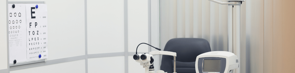
Advanced imaging using cutting-edge technology to capture high-resolution images is revolutionizing eye care. Beyond the basics of a comprehensive eye exam, ophthalmologists can advance their understanding of ocular conditions with a variety of testing options including optical coherence testing (OCT) that includes Swept-Source OCT, Spectral-Domain OCT and Optical Coherence Tomography Angiography, fundus photography, fundus autofluorescence and confocal microscopy. Optical coherence testing is a non-invasive imaging technique that uses light waves to capture cross-sectional 2D images within the structures of the eye such as the retina, optic nerve and cornea. Variations of OCT use either specialized light and lasers or detection mechanisms for faster image acquisition and higher resolution. Fundus photography takes detailed photographs of the back of the eye as well as the interior surface of the eye with a specialized camera that has a low-powered microscope and bright light source. Unlike fundus photography, fundus autofluorescence uses an external light to illuminate the eye to get detailed images to reveal patterns and variations in both intensity and distribution of the auto fluorescent signals across the retina. Confocal microscopy examines the structures of the eye at a cellular level detecting abnormalities in the cornea, conjunctiva and other ocular tissues that may impact corneal nerve density and be responsible for conditions like diabetic neuropathy and corneal neuropathic pain.
New trends in ophthalmology research as it relates to exploring advanced imaging in ophthalmology beyond the basics include artificial intelligence, drug development, fundus imaging technology and telemedicine. For the best services for your specific vision and eye health needs, schedule an appointment with Pennachio Eye by calling 325-227-1999.